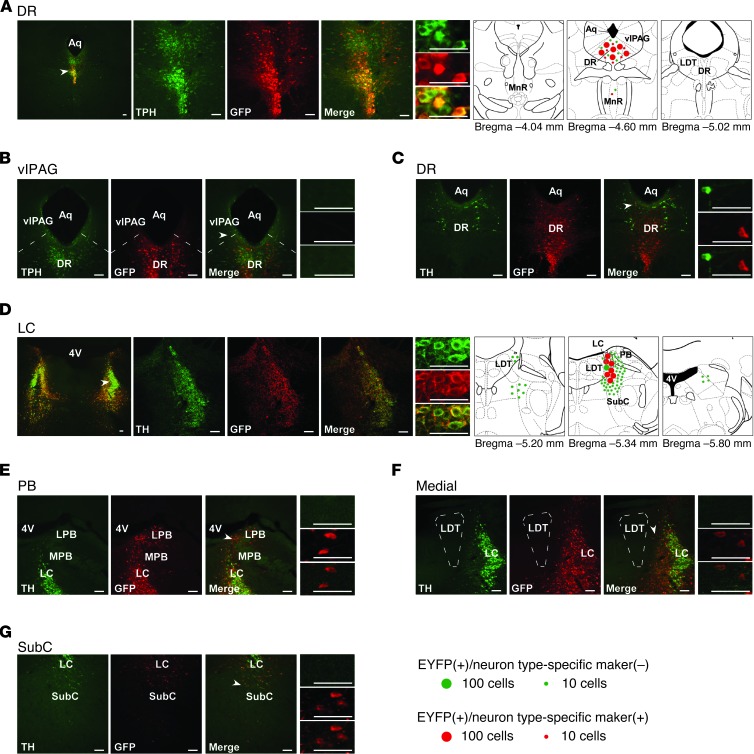
Figure 4. Restoration of orexin receptors in the DR or LC of Ox1r–/–Ox2r–/– mice using neuron type-selective promoters.
(A–C) Coronal brain sections from Ox1r–/–Ox2r–/–+5HT-OX2R mice (with targeted injection of AAV-Pet1/OX2R::EYFP) were double-stained with anti-GFP antibody (red) and either anti-TPH (A and B) or anti-TH (C, for dopaminergic neurons in the DR) antibody (green). (D–G) Sections from Ox1r–/–Ox2r–/–+NA-OX1R mice (with AAV-PRSx8/OX1R::EYFP injection) were double-stained with anti-GFP (red) and anti-TH (green) antibodies. Leaked expression of OX1R::EYFP in regions lateral (E, including PB), medial (F), and ventral (G, including SubC) to the LC (D) is shown. Regions denoted by white arrowheads are shown at higher magnification. Schematics show the spread of OX1R::EYFP or OX2R::EYFP expression. Mean numbers of EGFP+ cells are shown by green or red circles, indicative of the absence or presence, respectively, of the neuronal type–specific marker. For LC, only 1 side was shown. LPB, lateral PB; MPB, medial PB. Scale bars: 100 μm.

