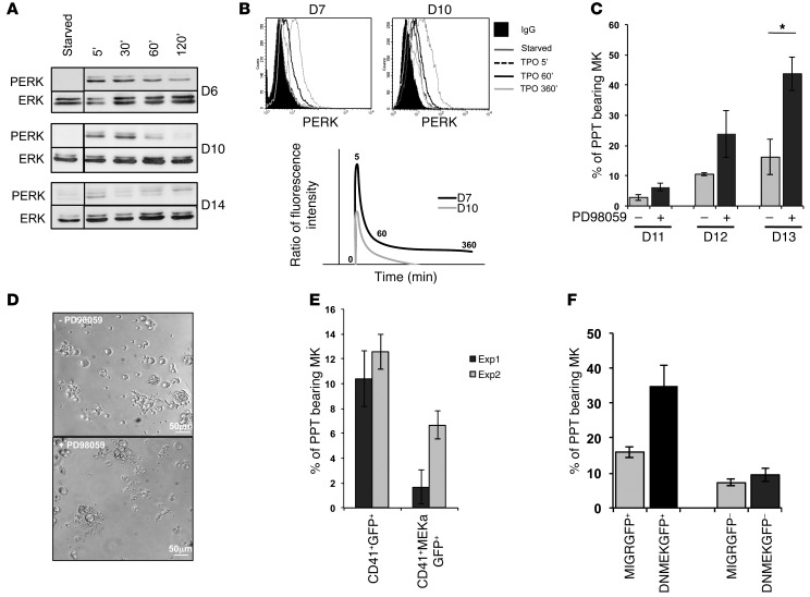
Figure 6. Decrease in MAPK signaling pathway is necessary for PPT formation by MKs.
MKs were differentiated from CD34+ cells in the presence of TPO. (A and B) Analysis of the MAPK pathway. Starved MKs were stimulated by TPO (100 ng/ml) for 5, 30, 60, and 120 minutes. (A) Western blot analysis of ERK and PERK performed at days 6, 10, and 14. (B) Flow cytometry analysis of PERK at days 7 and 10. The ratio of fluorescence intensity represents the ratio between the value obtained using PERK and control IgG isotype Abs for each time point of TPO stimulation. For A and B, experiments were performed 3 times with similar results. (C) Inhibition of MAPK pathway increases PPT formation. The CD41+ cells were sorted at day 6, the PD98059 inhibitor was added at day 8, and the percentage of PPT-bearing MKs was evaluated at days 11–13. One of 3 independent experiments with similar results is presented. Data were collected from triplicate wells and represent mean ± SD of triplicate. *P < 0.05, Student’s t test. (D) PPT-forming MKs cultured in presence or absence of PD98059 at day 13. (E and F) Inhibition or activation of MAPK pathway displayed an opposite effect on PPT formation. (E) CD34+ cells were transduced by retroviruses encoding for a dominant negative form of MEK1 and GFP (DNMEKGFP) or (F) for an active form of MEK1 (MEKa) and GFP. A retrovirus MIGR encoding for GFP was used as control. CD41+GFP+ cells were sorted at day 10 and the percentage of PPT-bearing MKs was evaluated at day 13 of culture. Data were collected from triplicate wells and represent mean ± SD.

