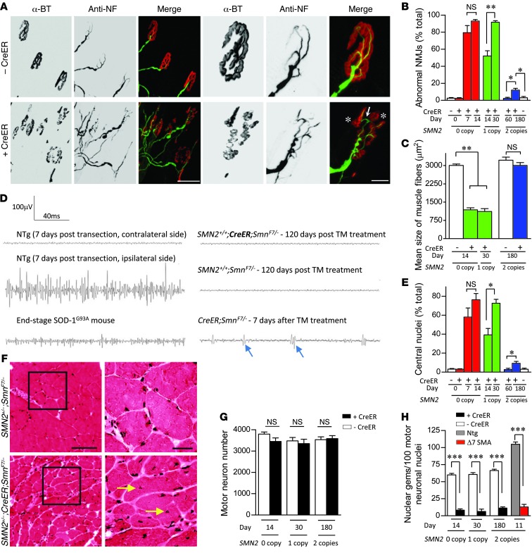
Figure 3. Age-dependent emergence of NMJ and muscle pathology in SMN-depleted mutants.
(A) Immunostains of NMJs in the gastrocnemius muscle of end-stage 0- or 1-copy SMN2 inducible mutants revealed profound abnormalities including AChR cluster fragmentation (asterisks) and imperfect overlap between pre- and postsynaptic regions (arrow). Depicted are NMJs from a TM-administered CreER;SmnF7/– mouse and a control without the CreER transgene. Scale bars: 100 μm; 30 μm (detail). (B) Quantification of NMJ defects in 0-, 1-, and 2-copy SMN2-inducible mutants. *P < 0.05; **P < 0.01. n = 300 NMJs from each of 3 mice of each genotype, 1-way ANOVA. (C) Normal muscle fiber size in the gastrocnemius of homozygous SMN2 but not hemizygous or 0-copy SMN2 inducible mice is indicative of relatively normal innervations in the presence of 2 SMN2 copies. **P < 0.01, 1-way ANOVA. n > 50 fibers from each of 3 or more mice of each genotype. (D) Representative EMG traces recorded from the gastrocnemius muscle of 2-copy SMN2-inducible mutants and relevant controls. No evidence of denervation was detected. Shown are fibrillation potentials (arrows), a sign of denervation in CreER;SmnF7/– mutants, end-stage SOD-1G93A ALS model mice and animals in which the sciatic nerve was transected. (E) Extent of centrally nucleated myofibers in the various cohorts of SMN-inducible mutants. Statistics calculated as in C. (F) H&E stains of gastrocnemius muscle in transverse section are illustrative of a primary myopathy in the SMN-inducible mice; arrows indicate central nuclei. Scale bars: 100 μm; 30 μm (inset). Quantification of (G) motor neurons and (H) motor neuronal gems failed to provide evidence of cell loss despite profound SMN depletion. Motor neurons and gems in each of 3 mice of the various genotypes were quantified. ***P < 0.001, t test.

