Figure 1.
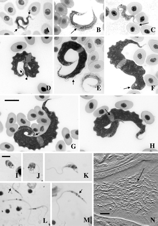
Brightfield images of Giemsa stained fish blood films (A-H) and leech squashes (I-M); differential interference contrast image of haematoxylin and eosin stained histological section through a leech (N). A: Small, likely dividing trypanosome with two kinetoplasts (arrows) from Clinus agilis at Mouille Point. B: Small trypanosome showing the nucleus (arrow) from Clinus cottoides at Koppie Alleen. C: Small trypanosome demonstrating the flagellum from Parablennius cornutus at Koppie Alleen. D: Large trypanosome with faintly stained flagellum (arrow) and showing the position of the kinetoplast (arrowhead), from Clinus superciliosus at Tsitsikamma. E: Large trypanosome (left) demonstrating the undulating membrane (arrow) and small form (right) from C. superciliosus at Tsitsikamma. F: Large form with bluntly rounded posterior (arrow) from C. superciliosus at Tsitsikamma. G: Large form with hooked posterior (arrow) from C. superciliosus at Tsitsikamma. H: Large form demonstrating striae (arrow) from Clinus superciliosus at Tsitsikamma. I: Amastigote, (J) sphaeromastigote, (K) short, thick epimastigote, (L) slender epimastigotes, one with two flagella (arrow), (M) slender epimastigote (arrow) with three nuclei and two kinetoplasts, all from Zeylanicobdella arugamensis from Koppie Alleen. N: Numerous long slender epimastigotes (arrow) in the dorsal sinus of an adult Z. arugamensis from Koppie Alleen. Scale bars: A-H = 10 μm; I-M = 5 μm; N = 20 μm.

