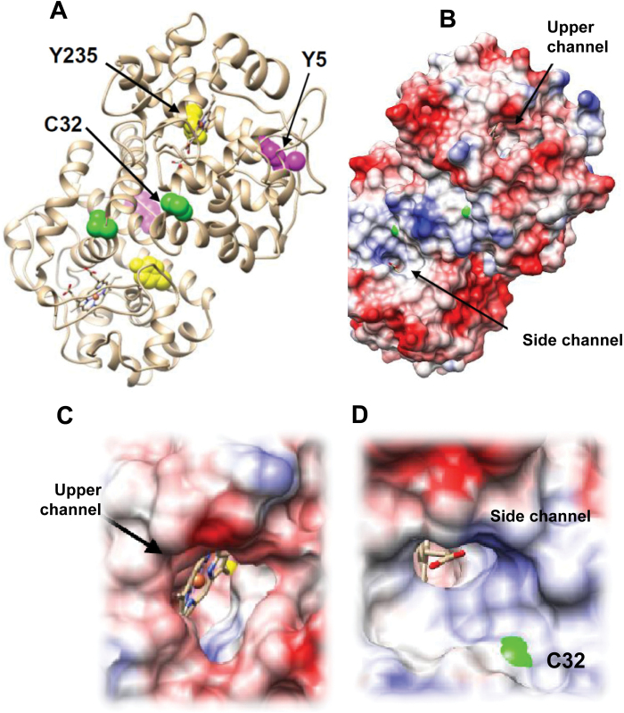
Fig. 4.
(A) Structure of homodimeric pea APX (PDB ID: 1apx). Residues identified as a target of tyrosine nitration and S-nitrosylation are shown as space filling. (B) The haem group is enclosed in a pocket with two channels to the exterior. (C) The view along the upper channel reveals that Y235 is at the bottom of the pocket. (D) C32 is located at the ascorbate binding site in the vicinity of the side channel. (This figure is available in colour at JXB online.)

