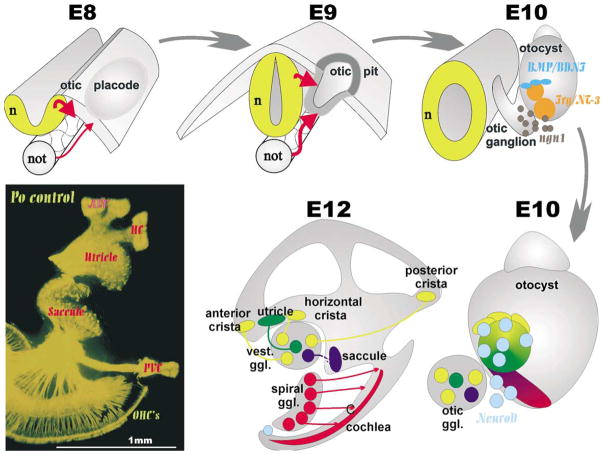
Fig. 1.
This diagram illustrates the origin of primary neurons in a mammalian otocyst. Inductive interactions transform ectoderm lateral to the developing hindbrain into the otic placode that invaginates to form the otocyst. In the otocyst there is upregulation of ngn1 in areas which also express other markers such as BMP-4, lunatic fringe, BDNF and NT-3. Upregulation of ngn1 results in formation of primary neuron precursors which delaminate and migrate to the forming otic ganglion. These primary neuron precursors upregulate another bHLH gene, NeuroD while they delaminate from areas that are close by or appear to become distinct sensory epithelia. Whether or not the vestibular primary neurons that project to a given sensory epithelium are actually delaminating from or nearby the sensory epithelia they later are connected to remains unknown. At birth (lower left) the innervation of the ear is distinct to all endorgans.

