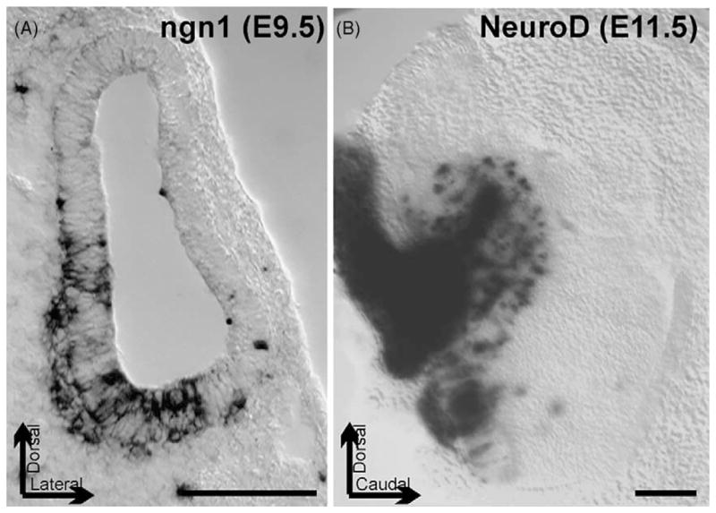
Fig. 2.

These images show the expression of the bHLH gene ngn1 in a 9.5-day-old mouse embryo otocyst as revealed by in situ hybridization, and of NeuroD in the developing cochlear and vestibular primary neurons of a 11.5-day-old mouse embryo using a LacZ reporter. Note that ngn1 is expressed inside the otocyst wall as well as in delaminating sensory neurons. NeuroD expression is both in the delaminated differentiating primary neurons that form the otic (vestibulocochlear) ganglion (left) and in cells in the otocyst wall. Bar indicates 100 μm, arrows indicate position of the preparations.
