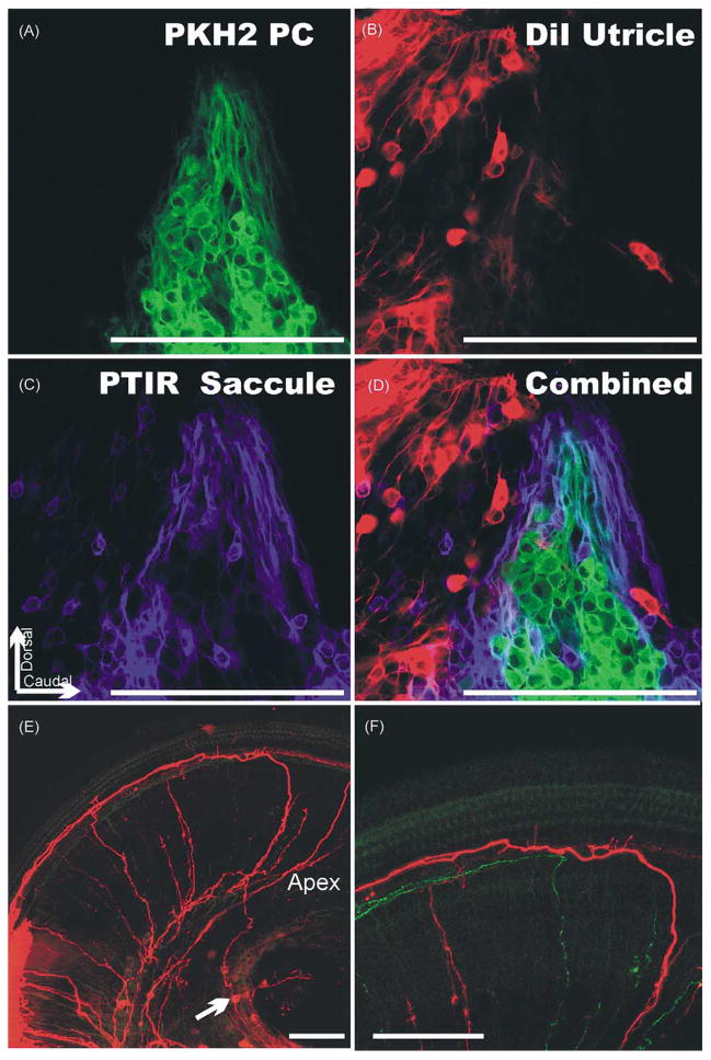
Fig. 4.
Three differently fluorescing lipophilic tracers were used to selectively label the vestibular primary neurons that project to the posterior vertical crista (PC; A), the utricle (B) and the saccule (C). The combination of all three shows there is segregation of PC primary neurons into a single group. Nevertheless, other primary neurons projecting to other endorgans are interspersed among them (D). Note also the complete absence of any double labeling (D) suggesting that each primary neuron projects to only one endorgan. The cochlear labeling (E, F) shows labeling of an apical spiral primary neuron (arrow in E) that projects first about 200 μm apical, runs in a radial bundle to the cochlea where it forms collaterals to inner hair cells (F) and extends for 500 μm towards the base where the fiber was labeled by the DiI injection (E). Bar indicates 100 μm.

