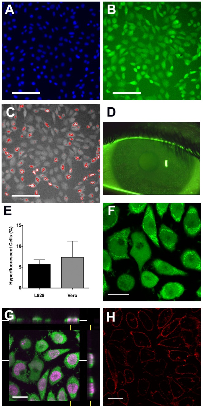
Figure 1. Fluorescein staining of control cell populations.

L929 cell cultures were treated with fluorescein and Hoescht 33342 prior to observation. Nuclear staining is shown in (A) and fluorescein staining in (B) with typical hyperfluorecent cells visible. Images were obtained using the ArrayScan®II system and cells categorized by fluorescein intensity with hyperfluorescent cells identified shown in (C). ‘Solution induced corneal staining’, as seen on a slit lamp biomicroscope, is shown (D) for comparison, in which characteristic hyperfluorescent punctate spots are readily apparent. The proportion of hyperfluorescent cells in L929 (n = 20) and Vero cultures (n = 6) was similar (E), with bars showing standard error. Confocal microscopic analysis of Draq5 (a nuclear stain) and fluorescein-stained L929 cells, revealed the presence of fluorescein throughout the interior of the cell, with numerous highly intense fluorescein-containing structures being visible in the cytoplasm, especially of hyperfluorescent cells (F shows a single confocal ‘slice’ through the cells, and (G) shows the orthogonal view; a 3D reconstruction of staining along the white and yellow axes). Treating control cells with the membrane-slective stain Vybrant ® DiI confirmed the likely appearance after staining with a compound, which localizes on the cell surface, providing further confirmation that fluorescein has entered cells (H). Data shown are representative of several experiments. Scale bars in (A) to (C) represent 100 µm and in (F) to (H) represent 20 µm.
