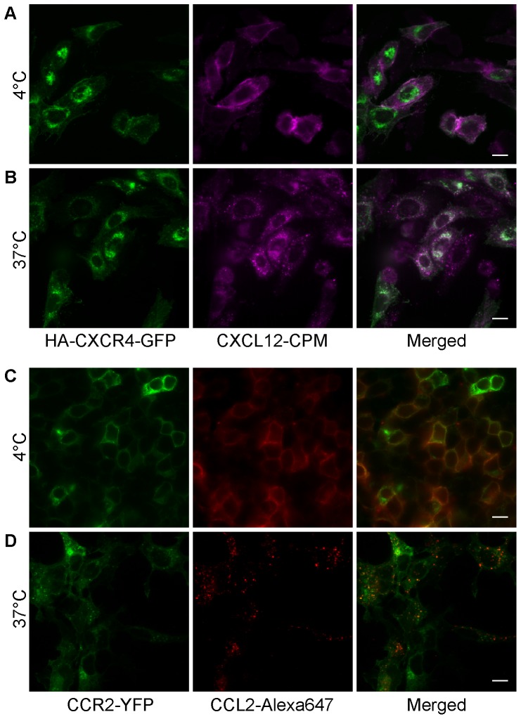
Figure 4. Microscopy imaging of chemokine-receptor interactions and chemokine-mediated receptor internalization.
(A) CHO-K1 cells transiently transfected with CXCR4-GFP were stained with 100 nM CXCL12-CPM at 4°C. Left, CXCR4-GFP fluorescence. Middle, CXCL12-CPM. Right, merged image. (B) CHO-K1 cells expressing CXCR4-GFP were incubated at 37°C for 30 min following the surface staining at 4°C with CXCl12-CPM. Left, CXCR4-GFP. Middle, CXCL12-CPM. Right, merged image. (C) Staining of HEK293t cells transiently expressing CCR2-YFP by CCL2-Alexa647 at 4°C. Left, CCR2-YFP fluorescence. Middle, CCL2-S6-Alexa647. Right, merged image. (D) HEK293t cells expressing CCR2-YFP were incubated at 37°C for 30 min following the surface-staining with CCL2-Alexa647 at 4°C. Left, CCR2-YFP. Middle, CCL2-Alexa647. Right, merged image. (Scale bar: 10 µm).

