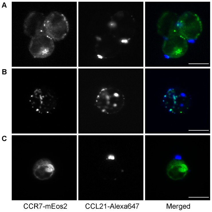
Figure 5. Microscopy imaging of CCL21 on CCR7 expressing cells using CCL21-Alexa647.
CHO-K1 cells expressing CCR7-mEos2 were stained in suspension for 30 min on ice with (A) 100 nM, (B) 50 nM, and (C) 25 nM CCL21-Alexa647. Left, CCR7-mEos2 fluorescence. Middle, CCL21-Alexa647. Right, merged image. (A) and (C) show that the puncta are clearly on the outside of the cell. (B) shows that occasionally multiple puncta are observed. (Scale bar: 10 µm).

