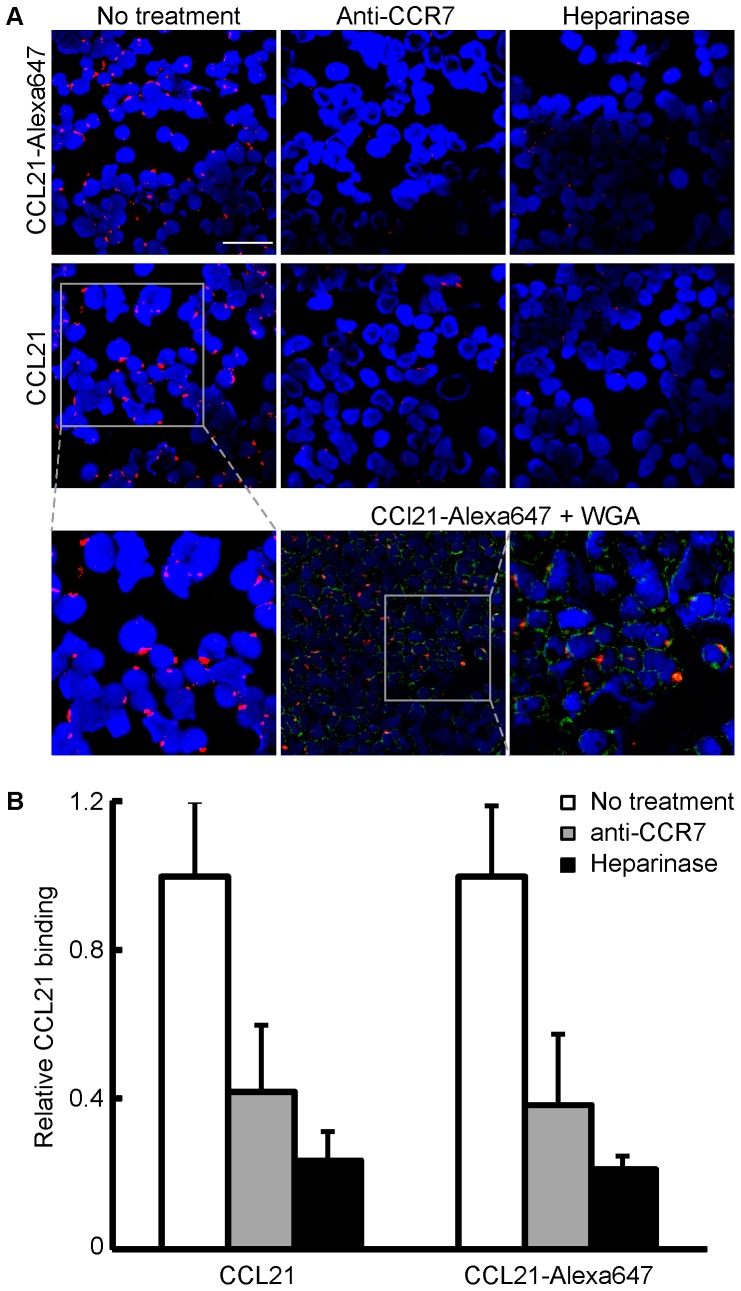
Figure 6. Microscopy imaging of CCL21 on CCR7 expressing cells by immunofluorescence.
(A) Binding patterns for both 647-labeled and unlabeled CCL21 (10 nM) show a punctate binding pattern on the surface of HEK293t cells transfected with CCR7. Labeled or unlabeled recombinant human CCL21 were incubated with HEK293t cells, and binding was detected by immunofluorescence staining. Red signal: CCL21; blue signal: DAPI nuclear stain; green signal: membrane staining by wheat germ agglutinin (WGA) conjugated to Alexa-fluor 488(Scale bar: 50 µm). (B) To assess the extent of interaction, the areas occupied by both CCL21 signal (Red) and DAPI (Blue) were quantified, and the CCL21/DAPI ratio in the untreated sample was defined as 1, and the relative binding in all other samples is expressed as its fraction.

