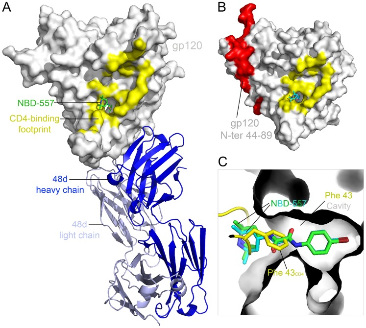
Figure 2. Structures of HIV-1 gp120 core in complex with NBD-557.
(A) YU2 gp120 coreV3s (surface representation in grey) in complex with NBD-557 (stick representation in green) and Fab 48d depicted in a ribbon diagram (light chain in light blue and heavy chain in blue). (B) NBD-557 (stick representation in cyan) binds the Phe 43CD4 cavity on clade A/E93TH057 gp120 coree. Area colored red represents N-terminal residues (44–89), which are missing in gp120 core in (A). CD4 footprints on gp120 are colored in yellow in (A) and (B). (C) Superposition of NBD-557- and Fab 48d- bound YU2 gp120, NBD-557-bound clade A/E93TH057 gp120 core, and the CD4-bound gp120. Two NBD-557 (green and cyan) and the side chain of Phe 43CD4 (yellow) in the cavity are highlighted.

