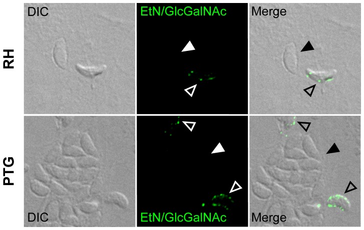
Figure 3. Protein-free Glc-GalNAc-substituted GPIs are clustered on extracellular parasites.
Staining after permeabilization of HFF cells infected (72 h p.i.) with RH (upper panel) and PTG (lower panel) strains was performed using mAb T54 E10, recognizing both the EtN-PO4 and the Glc-GalNAc side-branch epitopes of protein-free GPIs. Filled arrowheads point to intracellular parasites residing inside parasitophorous vacuoles. Unfilled arrowheads point to extracellular parasites. DIC, differential interference contrast.

