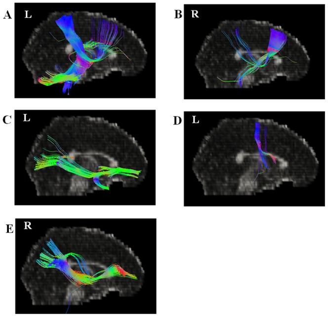
Figure 3. Fiber connectivity maps from starting points in all subjects.

(A) Arcuate fibers near left parietal lobe; (B) Cingulum near right corpus callosum-body; (C) Cingulum near left corpus callosum-genu; (D) Arcuate fibers near left precentral gyurs; (E) Inferior longitudinal fasciculus near right superior temporal gyrus.
