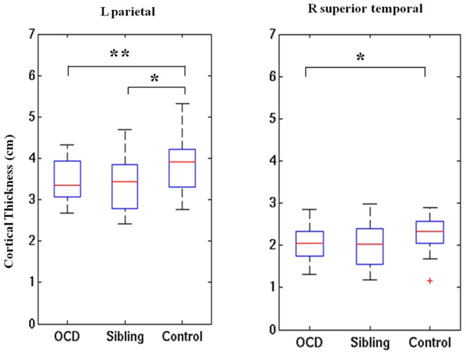
Figure 6. Significant group differences in cortical thickness in patients with OCD, their unaffected siblings, and healthy controls.

Arcuate fibers near left superior parietal lobule (on the left panel) and inferior longitudinal fasciculus near right superior temporal gyrus (on the right panel) were used as starting points, respectively. * P<0.05. ** P<0.01.
