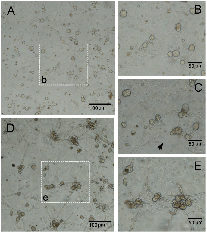
Figure 1. Culture development of locust frontal ganglion neurons into clustered networks.
(A) After 3 DIV, completely dissociated neurons had already started growing neuronal processes with continuous branching. The area outlined in (b) is enlarged in B. (C) Same area as in (B) but at 6 DIV. At this stage, neurons and small clusters of neurons are already densely connected and form a complex network. At the same stage, branched neurites (pointed by the black arrow) that failed to contact neighboring neurons start to retract. (D) Migration of neurons due to the tension along neurites leads to the formation of large neuronal clusters and of thicker bundles of neurites. For a better visualization, the area outlined in (e) is enlarged in E.

