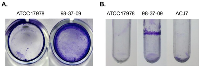
Figure 1. Polystyrene colonization.

Panel A. Representative images of A. baumannii strains ATCC17978 and 98-37-09 colonization of 24 well polystyrene microtiter plates as visualized by crystal violet staining after 48 h incubation at 37°C. Panel B. Representative images of A. baumannii strains ATCC17978, ACJ7, and 98-37-09 colonization of polystyrene tubes after 48 h incubation.
