
Figure 7.
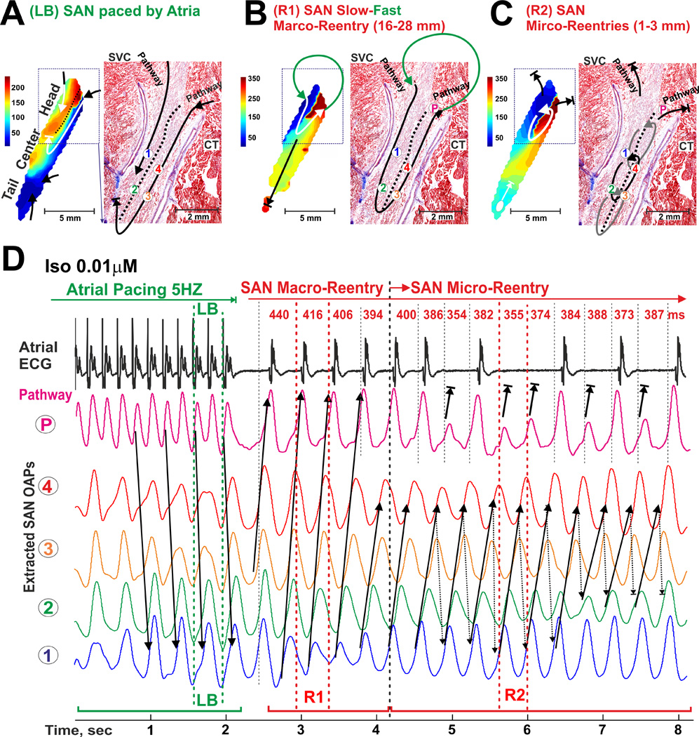
Induction of a counter-clockwise micro-reentry inside of the SAN during 0.01 µM Iso perfusion. (A) Longitudinal conduction block (LB) induced by atrial tachypacing (5 Hz) on the border of different refractoriness within the SAN (see Figure 5 for Iso 0.01 µM) is shown similar to Figure 6A. (B) SAN fast-slow macro-reentry (R1) through the SAN occurred in the MI canine heart #3 within the 10 s after cassation of atrial tachypacing is shown (see also Movie 4). (C) SAN slow-slow micro-reentry (R2) transformed from the previous macro-reentry is shown. (D) OAPs selected from the right superior SACP (P) and the SAN micro-reentry circuit (1–4) trace LB formation as well as macro- and micro-reentry formation and spontaneous termination. Size and location of the reentrant circuits were determined by histological examination. During different reentry episodes (1–8 sec), the atrial myocardium was activated via the same SAN conduction pathway as in normal sinus rhythm. Abbreviations are the same as those in Figure 2.