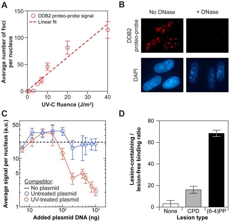
Figure 2. The DDB2 proteo-probe recognizes 6-4-photoproducts in vitro.
(A) The DDB2 proteo-probe signal increases linearly with fluence (J/m2). Fibroblasts were irradiated with different doses of UV-C. Each point is an average of three replicas. Each replica represents an average of at least 60 cells. Dashed line: linear fit (R2 = 0.94). Error bars: s.e.m. (B) The DDB2 proteo-probe signal is DNA-dependent. Fibroblasts were irradiated with UV-C (10 J/m2), and untreated or treated with DNase. Nuclei are visualized by DAPI staining. (C) The DDB2 proteo-probe signal can be competed with UV-treated plasmid DNA. Fibroblasts and plasmid DNA were irradiated with UV-C (10 J/m2 and 300 J/m2, respectively). The DDB2 proteo-probe was incubated with plasmid DNA prior to hybridization onto irradiated fibroblasts. Dashed line: no plasmid control proteo-probe signal level. Each point is an average of three replicas. Each replica represents an average of at least 400 cells. Error bars: s.e.m. (D) The DDB2 proteo-probe binds preferentially to 6-4-photoproducts [(6-4)PP] over cyclobutane pyrimidine dimers (CPD). The DDB2 proteo-probe was immobilized on agarose beads, and incubated with the DNA restriction fragments of a plasmid containing, or not, a unique lesion [(6-4)PP or CPD]. The average ratio of the amount of lesion-containing over lesion-free DNA fragments bound to the proteo-probe is shown (n = 3). Error bars: s.e.m.

