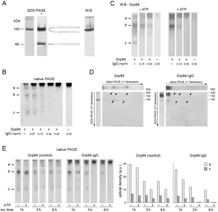
Figure 1. Characteristics of the complex formed by native rat Grp94 with IgG in native conditions.
(A) A representative fraction of peak 2 from gel filtration was analyzed in SDS-PAGE (8.5% polyacrylamide gel), in both non-reducing and reducing conditions (*) of samples (3 µg proteins/lane). Mass analysis (MALDI-TOF-TOF) for amino-acid sequence determinations was applied to bands (resolved in SDS-PAGE in reducing conditions) after controlled tryptic digestion. The more frequent peptide sequences of each band are indicated on right of the lane, whereas Western blotting (monoclonal anti-Grp94 Abs) is performed on the same fraction following SDS-PAGE in non-reducing conditions. (B) Native Grp94 of peak 2 (0.1 mg/ml, 10 mM Tris, pH 7.0) was incubated at 37°C for 120 min in absence of ATP, both alone (control) and with IgG at the indicated concentrations. Three µg of Grp94 were analyzed in native PAGE (8% acrylamide, pH 8.0) and gels stained with Coomassie blue. Lane on right, control IgG at the highest concentration. a, b and c: Grp94 bands with increasing electrophoretic mobility. (C) Western blotting for Grp94 on samples as in (B) processed in native PAGE in both absence and presence of ATP (1 mM) preincubated with Grp94 for 15 min before the addition of IgG. (D) 2D-PAGE of Grp94 alone (control) and with IgG (at the Grp94∶IgG molar ratio of 1∶2) after incubation at 37°C for 120 min in the presence of ATP, following native PAGE of samples, as in (B) and (C). The lanes of Grp94 alone (3 µg) and with IgG were cut and submitted to the second dimension on 10% acrylamide gel (see Methods). Above and on the left of each gel are lanes of reference of the first dimension and of SDS-PAGE (in non-reducing conditions), respectively. Long arrows indicate the direction of the run (from the cathode to the anode) in both PAGEs, whereas short arrows in the gels mark Grp94 bands that disappear after co-incubation with IgG (gel on right). (E) Native PAGE of Grp94 incubated with 0.3 mg/ml IgG and processed as in B at the indicated incubation times, in both absence and presence of ATP (as in C). On right: histograms representing the densitometric analysis of bands b and c of each lane (Gel-Pro Analyzer software, version 3.1). Heights of histograms indicate the optical density (in arbitrary units).

