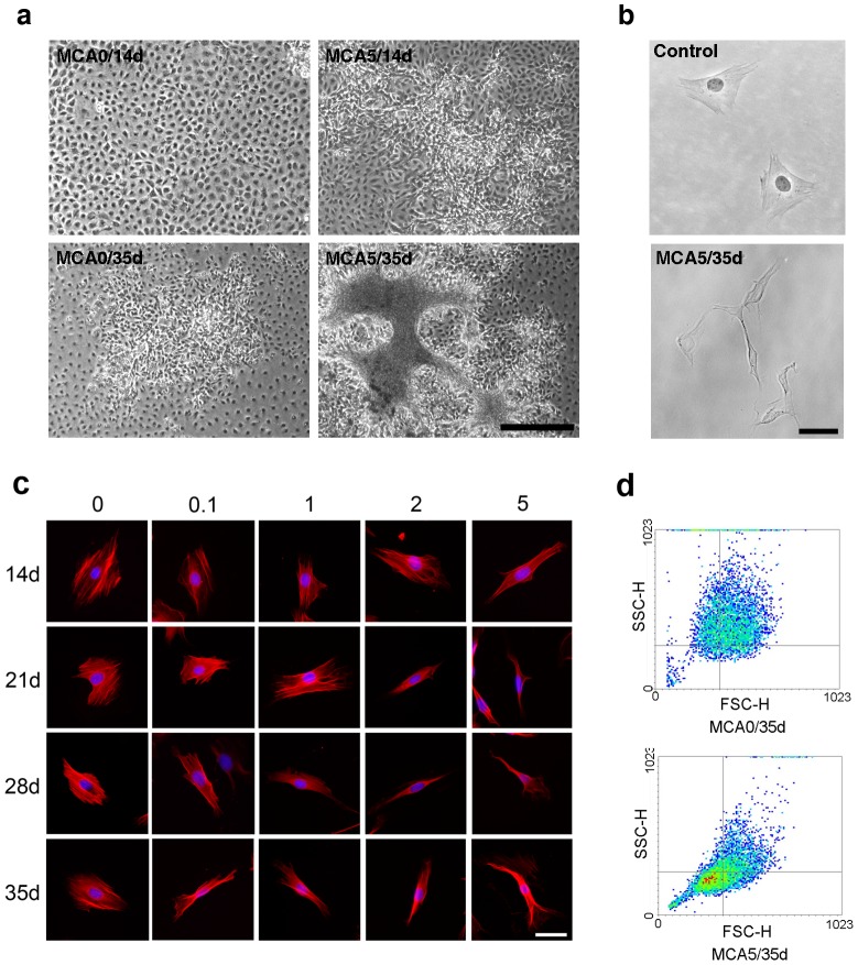
Figure 1. MCA induced cell transformation of BALB/c 3T3 cells.
(a) Representative microscopic appearance of MCA-induced foci. BALB/c 3T3 cells were treated with 5 μg/ml MCA (MCA5) and vehicle (MCA0) separately, and cultured for 14 (14d) and 35 days (35d); scale bar = 500 μm. (b) Cell morphology of individual cells of untreated BALB/c 3T3 cells (control) and MCA-treated BALB/c 3T3 cells (MCA5/35d); scale bar = 50 μm. (c) Alteration of morphological phenotypes and cytoskeleton organization of BALB/c 3T3 cells treated by different MCA doses (0.1, 1, 2, 5 μg/ml) and cultured periods (14, 21, 28, 35 days); smooth muscle actin (red), DAPI (blue), scale bar = 50 μm. (d) Cells in the control and MCA-treated groups were analyzed using flow cytometry. The forward scatter (FSC) and side scatter (SSC) of particles were simultaneously measured. All experiments had been repeated at least three times.

