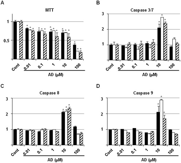
Figure 1.

Optimal dosage of AD used as an apoptosis-inducible positive control. RAW264.7 (closed box), MC3T3E1 (open box) and ATDC5 (shaded box) cells were seeded at a density of 1×10⁴ cells/well in 96-well tissue culture plates, and grown in the absence (Cont) or presence of 0.01 to 100 μM of AD. After 24 h, cells were subjected to MTT (A), and caspase 3/7 (B), 8 (C) and 9 (D) assays. Data are means ± standard deviation of the values from 3 sets of cultures. *, p < 0.05 vs each control.
