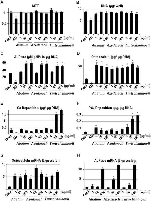
Figure 6.

Biochemical analysis and gene expression of OB-specific phenotypes induced by herbal extracts. A, MC3T3E1 cells were seeded in 96-well cell culture plates and allowed to differentiate into mature OBs. Then, cells were then cultured in the absence (Control) or presence of 10 μM AD, or 1, 10 and 100 μg/ml C. atratum (Atratum) M. azedarach (Azedarach) and C. turtschaninovii (Turtschaninovii) extracts. After 3 days, the cells were subjected to MTT assay. B, C, D, E and F, MC3T3E1 cells were seeded in 24-well cell culture plates and allowed to differentiate into mature OBs. Thereafter, cells were cultured in the absence (Control) or presence of 10 μM AD, or 1, 10 and 100 μg/ml C. atratum (Atratum) M. azedarach (Azedarach) and C. turtschaninovii (Turtschaninovii) extracts. After 7 days, the cell layer was lysed with 300 μl of 0.02% Triton-X 100 in saline. The aliquot was then subjected to DNA (B), ALPase activity (C), Ca (E), PO4(F) measurement. The CM was also concentrated and subjected to ELISA for osteocalcin (D). Data are means ± standard deviation of 3 sets of cultures and each value (except for DNA) is standardized to DNA content (μg) in the cell layer. G and H, MC3T3E1 cells were seeded and cultured, as described above. After 7 days, total cellular RNA was purified, and real-time RT-PCR for osteocalcin (G) and ALPase (H) was carried out. Data are means ± standard deviation of 3 sets of cultures and each value is standardized to β-actin. *, p < 0.05 vs control.
