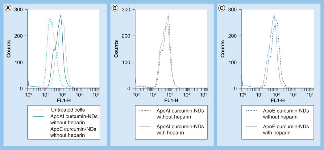
Figure 5. Effect of heparin on nanodisk-mediated curcumin uptake by SF-767 cells.
Cells were seeded at 1 × 10⁶ cells/well and incubated with curcumin NDs (20 µM curcumin) in the presence and absence of 50 µg heparin. After 1 h, cells were washed with phosphate-buffered saline and fresh medium was added. Cells were incubated further for 24 h and subjected to flow cytometry to detect internalized curcumin. (A) Untreated cells, ApoAI curcumin NDs without heparin and ApoE curcumin NDs without heparin. (B) Cells incubated with ApoAI curcumin NDs in the asbence and presence of heparin. (C) Cells incubated with ApoE curcumin NDs in the absence or presence of heparin. Data are representative of an experiment that was performed on two separate occasions. FL1-H: Height of the histogram that represents the mean curcumin fluorescence intensity detected by channel 1; ND: Nanodisk.

