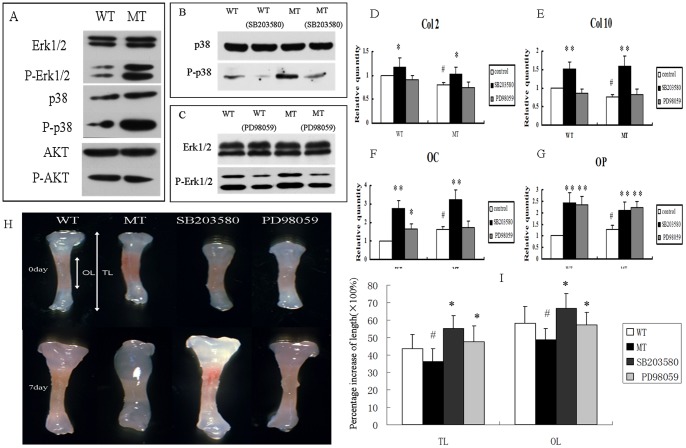
Figure 6. p38 and Erk1/2 pathway participated in the regulation of BMSCs by FGFR2.
(A) Western blotting demonstrated that levels of phospho-Erk1/2 and phospho-p38 were both increased in cultured BMSCs from MT mice. There was, however, no change in phospho-AKT. (B, C) The p38 and Erk1/2 pathways were inhibited by SB203580 and PD98059, respectively. (D–G) Relative expression of genes in BMSCs treated with SB203580 or PD98059 for 21 days. The expression levels of Col2, Col10, OC, and OP were significantly increased in both WT and MT BMSCs treated with SB203580; however, only OC and OP were increased after PD98059 treatment. (Student's t-test, *P<0.05, **P<0.01 versus untreated BMSCs, # P<0.05 versus wild-type BMSCs.). (H) Inhibition of p38 and Erk1/2 pathway by SB203580 and PD98059 rescued the growth retardation in cultured mutant tibia bones. The white double-headed arrows represent the total length (TL) and ossified tissue length (OL). (I) Percentage increases in TL and OL of phalange bones after culture for 7 days. (Student's t-test, #P<0.05 versus wild-type *P<0.05 versus mutant type.)

