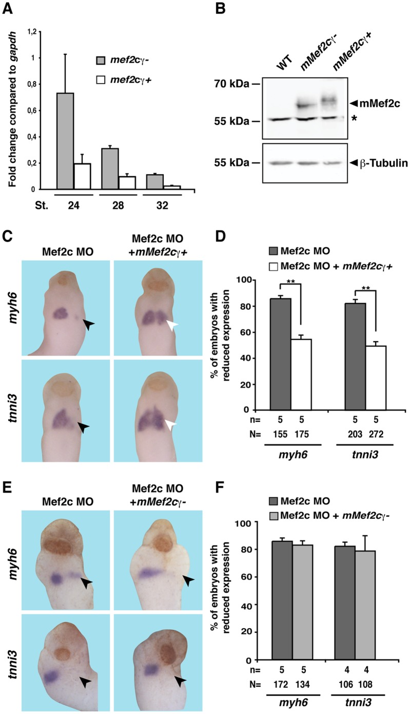
Figure 5. Specificity of phenotype observed upon knock down of Mef2c.
A. mef2cγ- and mef2cγ+ isoforms are expressed on RNA level in heart tissue enriched explants at stages 24, 28, and 32 as revealed by qPCR. Expression is shown relative to gapdh. B. mMef2cγ- and mMef2cγ+ are expressed on protein level upon RNA injection into Xenopus embryo at comparable levels. β-Tubulin served as loading control. Note that the Mef2c antibody used does not recognize endogenous Xenopus Mef2c protein. The asterisk indicates unspecific background. C–F. Mef2c MO was unilaterally injected together with GFP, mMef2cγ+ or mMef2γ- RNA as indicated. C, E Expression of the cardiac marker genes myh6 and tnni3 was monitored at stage 20 or stage 28. Black arrowheads indicate reduced marker gene expression, white arrowheads highlight the rescued situation. Ventral views of embryos are shown. D, F. Quantitative presentations are shown. N: number of examined embryos; n: number of independent experiments; st: stage; **, p≤0.01.

