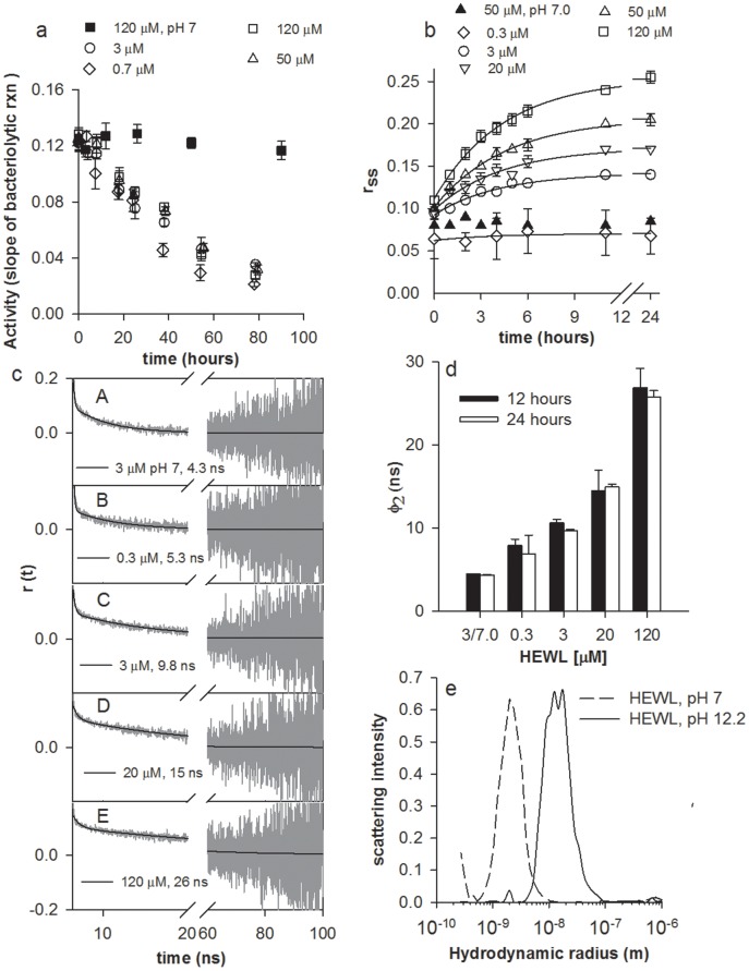
Figure 2. Activity and size of HEWL aggregates.
a, Bacteriolytic activity (in ▵Abs/min) of HEWL (diluted to 70 nM in assay cuvette) measured at pH 7 with pppppppwith M. lysodeikticus is highlighted at different time intervals subsequent to incubation of HEWL at concentrations 0.7, 3, 50 & 120 µM in pH 12.2 and 120 µM in pH 7. b, Changes in steady state fluorescence anisotropy (rss) of dansyl-HEWL conjugates at different time intervals in presence of 0.3, 3, 20, 50 & 120 µM of HEWL incubated in pH 12.2 and 50 µM in pH 7 is shown along with fitted curve for pH 12.2 (Eq. 2, Table 1). c, Nanosecond time-resolved fluorescence anisotropy decay of dansyl-HEWL conjugates after 24 hours for following protein concentrations (pH): A: 3 µM (7); B—E: 0.3, 3, 20 and 120 µM (12.2). The dark continuous line depicts the fit and extracted value for φ2 using tail fit analysis (Table S1). d, Average global rotational time (φ2) for 0.3—120 µM HEWL at pH 12.2 and 3 µM at pH 7 (extreme left) after 12 & 24 hours (Table S2). e, Size of HEWL aggregates revealed by dynamic light scattering after incubation of 120 µM HEWL in pH 12.2 (continuous line) and pH 7 (dashed line) for 26 hours. Peaks at Rh ∼2.0 nm indicate HEWL monomer, while those at 12.5 and 17.4 nm along with shoulders at Rh ∼9.0 and 28.7 nm reveal multimeric aggregates. Error bars, SD; N = 3.

