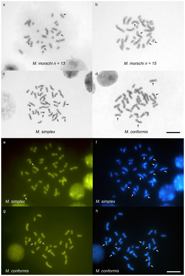
Figure 3. Mycetophylax metaphases submitted to C–banding technique and stained with fluorochromes.
(a) C – banding in the worker metaphase of M. morschi 2n = 26, (b) M. morschi 2n = 30, (c) M. simplex, (d) M. conformis denoting the heterochromatic positive bands (dark grey as indicated by the arrows). Metaphase of (e, f) M. simplex and (g, h) M. conformis stained with fluorochromes CMA3 and DAPI, respectively. The white arrows indicate the positive staining for CMA3. DAPI positivity was in agreement with the C – banding pattern (some positive bands are indicated by white arrows). Bar = 5 µm.

