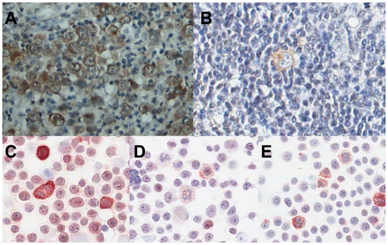
Figure 1. IGF-1R expression in HRS cells.
A, Representative cHL case showing expression of IGF-1R in the vast majority of HRS cells. B, pIGF-1R positive HRS cell. C, IGF-1R expression in L428, showing positive staining in all cells with strong expression in mitotic cells, D, IGF-1R expression in L1236, showing weak staining in all cells and strong expression in mitotic cells and, E, IGF-1R expression in KM-H2, showing weak staining with more pronounced expression in mitotic cells.

