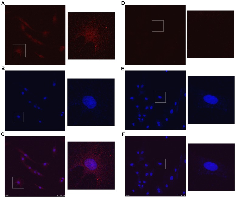
Figure 1. Confocal laser images of Kca3.1 channels in mesangial cell.
(A) the cytoplasm image stained by the anti-Kca3.1 primary antibody and the CyTm3-conjugated Affinipure Goat Anti-Rabbit lgG (H+L) secondary antibody, (B) the nucleus image stained by DAPI and (C) the image overlaid by A and B. Correspondingly (D), (E), and (F) are the images of controls without the primary antibody.

