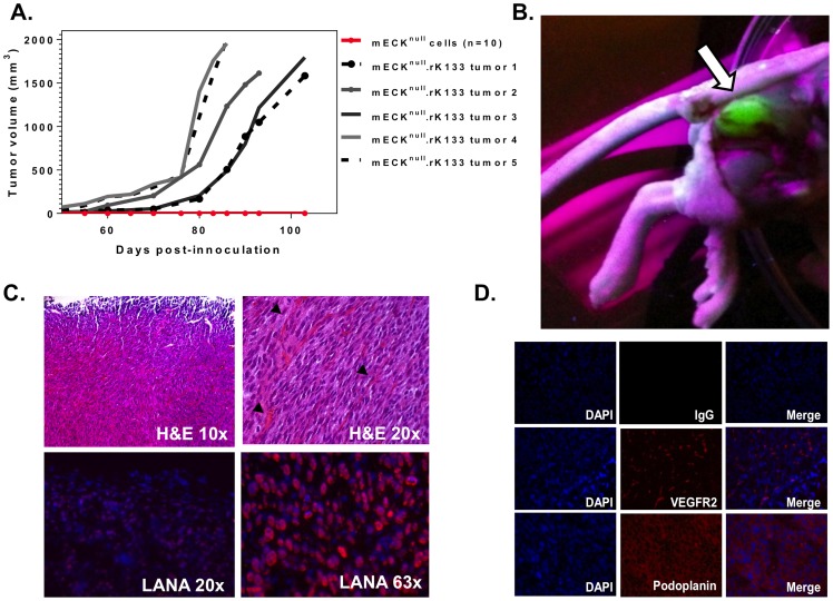
Figure 4. mECKnull. rK133 cells consistently form KS-like tumors in immunocompromised mice.
(A) Graph depicting tumor kinetics in athymic nu/nu mice. 3×10⁶ mECKnull. rK133 cells were subcutaneously injected into the hind flanks of 5 athymic nu/nu mice. Concurrently, 3×10⁶ mECKnull cells were subcutaneously injected into another group of 10 athymic nu/nu mice. Within 4–6 weeks solid mECKnull. rK133 tumors were palpable and growth was monitored by caliper measurements. mECKnull cells did not form tumors (red line, x-axis). (B) Dissection site showing a GFP+ tumor indicating the presence of rKSHV.219. The subcutaneous tumor was visualized under UV. (C) Tumor morphology was analyzed by H&E staining of paraffin embedded sections. Pathologically, they are composed of spindle cells arranged in bundles with RBCs in slit-like vasculature (top panels, black arrows). Frozen sections were prepared for immunofluorescence for KSHV LANA which showed that the spindle cells of the tumor express the viral latent nuclear antigen, LANA (bottom panels). (D) Immunofluorescence analysis of angiogenic protein expression in vivo reveals that the tumor cells express VEGF-R2 and podoplanin. A representative tumor is shown.

