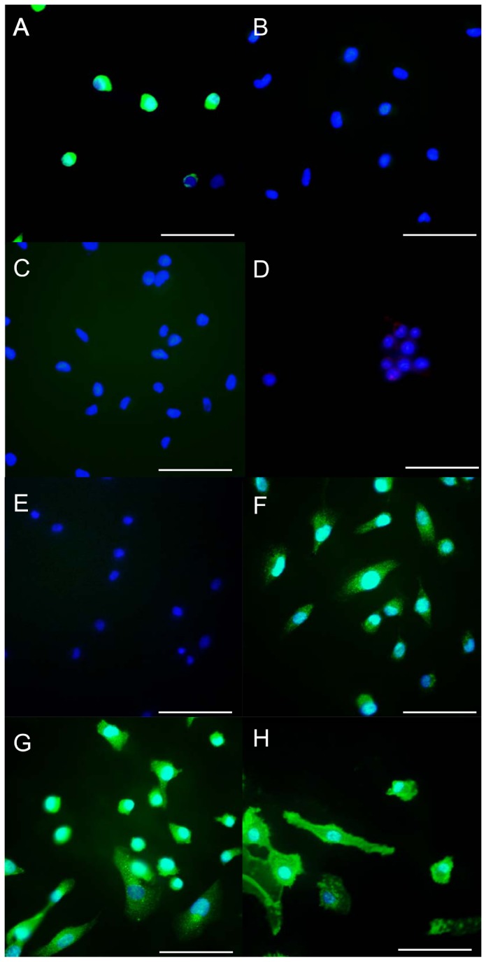
Figure 3. The results of the immunohistochemical analysis of the SCLs.

The SCLs were positive for vimentin (A), MMP-14 (F), MMP-2 (G) and CD44 (H). In contrast, the staining for α-SMA (B), VWF (C), CD31 (D) and desmin (E) was negative. (Scale bars show 50 µm.)
