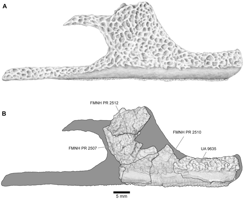
Figure 13. Reconstructions of right maxilla in labial view.
A, illustrated reconstruction based on composite digital model; B, outline reconstruction showing main specimens (FMNH PR 2507, FMNH PR 2510 [reversed], FMNH PR 2512 [reversed], UA 9635) in combined digital model. Additional data taken from neighbouring elements and positional information in skull reconstruction. See Figs 14, 15 for detailed views of individual specimens.

