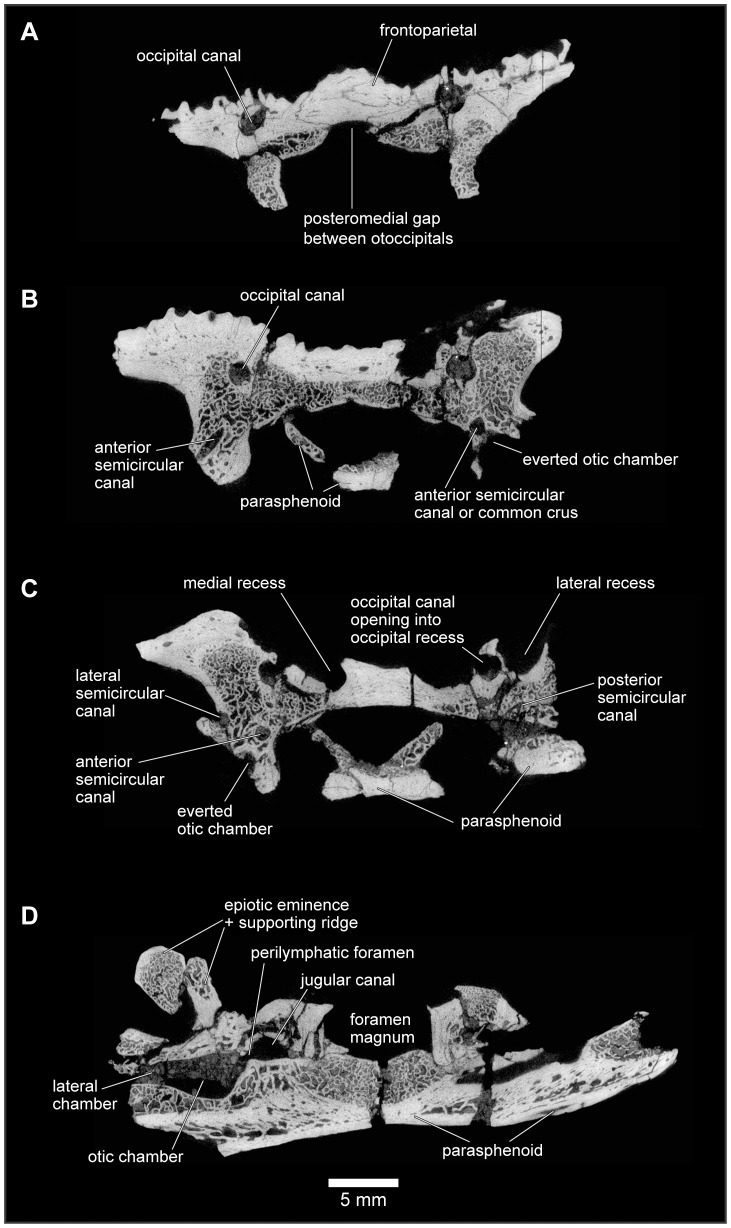
Figure 24. Approximately anterodorsal-posteroventral progression of μCT slices through braincase, FMNH PR 2512.
A, slice YZ 386; B, slice YZ 637; C, slice YZ 719; and D, slice YZ 992. Scan slices in YZ plane of reconstructed volume. Note that anteroposterior and dorsoventral biological axes deviate approximately 45° from scan reconstruction XZ and YZ axes.

