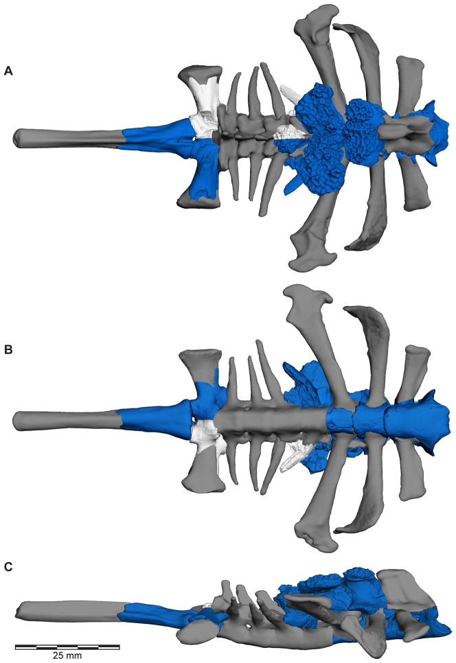
Figure 32. Three-dimensional digital reconstruction of axial skeleton of Beelzebufo ampinga.
A, dorsal; B, ventral; and C, right lateral views of axial column. As in Fig. 1, with material of Beelzebufo ampinga in dark blue. Mirrored left portion of neural arch of fifth presacral vertebra in model (FMNH PR 2512 Vertebra B) and centrum and transverse process of sacral vertebra (FMNH PR 2003) are mirrored in light grey. Dark grey postcranial elements modelled on large female specimen of Ceratophrys aurita (LACM 163430). See Supporting Information S1 for detailed description of model.

