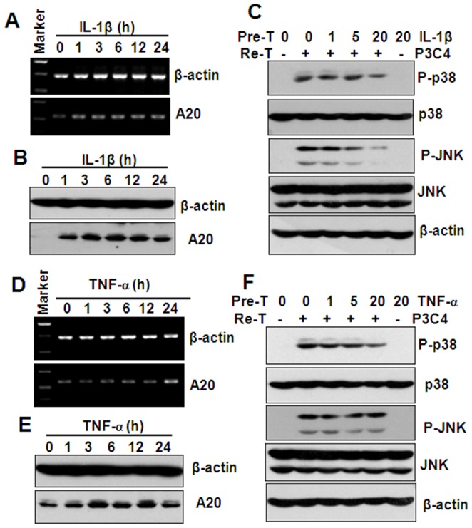
Figure 6. The effect of IL-1β and TNF-α pre-treatment on Pam3CSK4 (P3C4)-induced A20 expression and p38, JNK phosphorylation.

(A) RT-PCR analysis of A20 gene expression in THP-1 cells, treated with 20 ng/ml IL-1β for the indicated hours. β-actin gene expression was detected as loading controls. (B) Western blot analysis of A20 protein expression in THP-1 cells, treated with 20 ng/ml IL-1β for the indicated hours. β-actin protein was detected as loading controls. (C) Western blot analysis of p38, and JNK phosphorylation in THP-1 cells, pre-treated (Pre-T) with the indicated concentrations of IL-1β for 24 h, and re-treated (Re-T) with medium, or 1 µg/ml Pam3CSK4 for 30 min. β-actin protein was detected as loading controls. (D) RT-PCR analysis of A20 gene expression in THP-1 cells, treated with 20 ng/ml TNF-α for the indicated hours. β-actin gene expression was detected as loading controls. (E) Western blot analysis of A20 protein expression in THP-1 cells, treated with 20 ng/ml TNF-α for the indicated hours. β-actin protein was detected as loading controls. (F) Western blot analysis of p38, and JNK phosphorylation in THP-1 cells, pre-treated with the indicated concentrations of TNF-α for 24 h, and re-treated with medium, or 1 µg/ml Pam3CSK4 for 30 min. β-actin protein was detected as loading controls.
