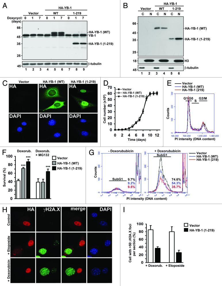
Figure 1. Effects of full-length and truncated YB-1 proteins on proliferation and survival during DNA damaging stress. (A) Expression of HA-tagged full-length and truncated YB-1 proteins in stably transfected NIH3T3 cells was induced by doxycycline. Whole-cell extracts were analyzed by WB using YB-1 antibodies. β-tubulin was used as loading control. (B) Nucleocytoplasmic distribution of HA-YB-1 (WT) and HA-YB-1 (1–219) was analyzed in cytoplasmic (C) and nuclear (N) fractions by WB using HA antibodies. Equal loading and fraction purity were controlled using Histone 3 and β-tubulin antibodies. (C) Subcellular localization of full-length and truncated YB-1 in NIH3T3 cells was examined by IF using HA antibodies. DAPI was used to visualize nuclei. (D) Growth curves of NIH3T3 cells expressing full-length or truncated YB-1. (E) Effect of YB-1 (WT) and YB-1 (1–219) expression in NIH3T3 cells on cell cycle progression. (F) Cells expressing full-length or truncated YB-1 were treated with 5 µM doxorubicin in the absence or presence of 10 µM MG132 for 14 h. Cell viability was determined by trypan blue staining (t test, P value [***] < 0.001). (G) Survival of YB-1 expressing NIH3T3 cells treated with 5 μM doxorubicin was monitored by FACS using PI staining. (H–I) NIH3T3 cells were transiently transfected with HA-YB-1 (1–219) and treated with 100 µM etoposide or 5 µM doxorubicin for 14 h and analyzed by IF using HA and γH2A.X antibodies, as indicated. DAPI was used to visualize nuclei (H). Percentage of cells containing more than 50 γH2A.X foci per section was determined by counting of >100 cells per field in 5 fields (I).

An official website of the United States government
Here's how you know
Official websites use .gov
A
.gov website belongs to an official
government organization in the United States.
Secure .gov websites use HTTPS
A lock (
) or https:// means you've safely
connected to the .gov website. Share sensitive
information only on official, secure websites.
