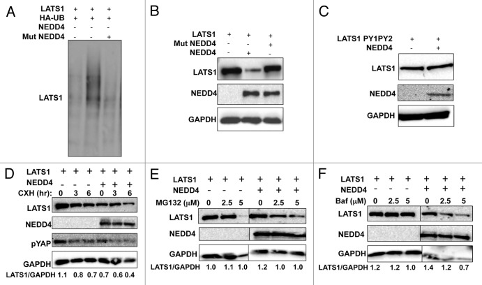
Figure 2. NEDD4 induces ubiquitination and affects the steady-state and half-life of LATS1 protein. (A) NEDD4 ubiquitinates LATS1 in vivo. HEK293T cells were transfected with the indicated plasmids. After 24 h, cells were treated with 20 μmol/L MG132 for 4 h. Lysates were prepared (B) and IP with anti-Max and detected with anti-HA antibodies (UB). (B and C) LATS1 steady-state stability is controlled by NEDD4. HEK293T cells were transiently transfected with Max-LATS1 or LATS1PY1PY2 (C) and NEDD4. After 24 h, cells were lysed and blotted as indicated. (D) ITCH reduces the half-life of LATS1. HEK293 cells were cotransfected with the indicated plasmids. After 24 h, cells were treated with 20 μg/mL cycloheximide (CHX) at the indicated time points and analyzed as shown in the figure. (E and F) LATS1 stability is controlled by the proteasomal pathway. HEK293T cells were transiently transfected with Max-LATS1 and NEDD4. After 24 h cells were treated with MG-132 (E), or bafilomycin [BAF] (F) as indicated for 4 additional hours. Equal amounts of total lysates were blotted as indicated. GAPDH, glyceraldehyde-3-phosphate dehydrogenase was always used as a protein loading control. Numbers under the blots represent the relative expression of LATS1 protein after correction with the loading control GAPDH expression levels.

An official website of the United States government
Here's how you know
Official websites use .gov
A
.gov website belongs to an official
government organization in the United States.
Secure .gov websites use HTTPS
A lock (
) or https:// means you've safely
connected to the .gov website. Share sensitive
information only on official, secure websites.
