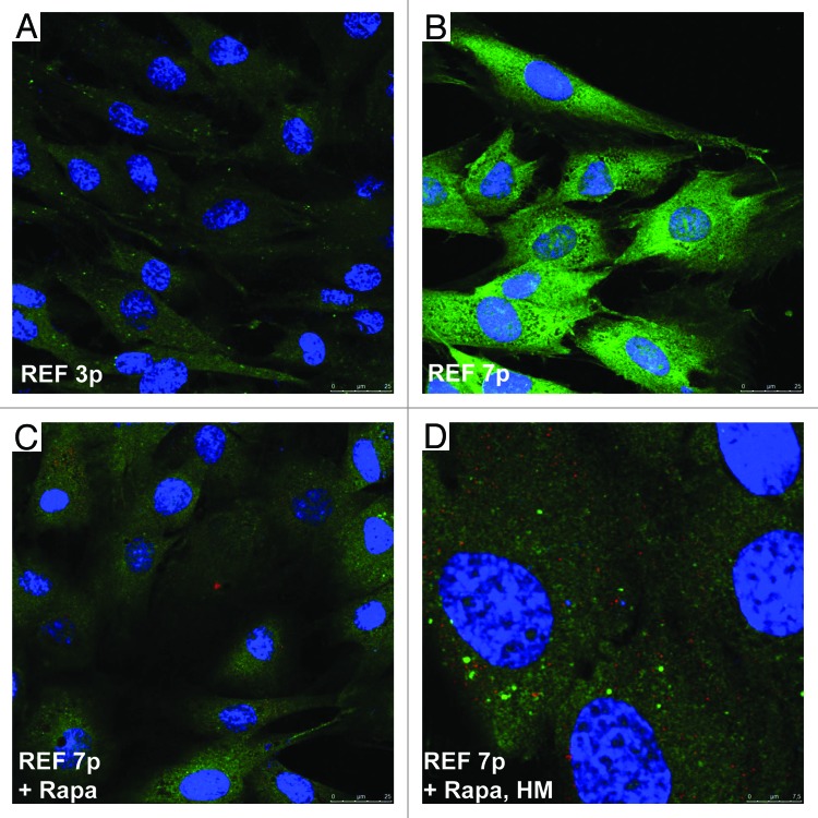
Figure 5. Immunofluorescence of LC3/LAMP1 staining in senescent REFs and rapamycin-derived cells. Confocal microscopy images were taken from rapamycin-untreated senescent REF cells (A) passage 3, (B) passage7; (Cand D) REF cells of passage 7 after short-term rapamycin treatment for 5 h (C, magnification as in A and B) and a higher magnification (D). Coverslips were stained with LC3 rabbit polyclonal and anti-LAMP1 mouse monoclonal antibody (Cell Signaling). Nuclei were stained with DAPI.

An official website of the United States government
Here's how you know
Official websites use .gov
A
.gov website belongs to an official
government organization in the United States.
Secure .gov websites use HTTPS
A lock (
) or https:// means you've safely
connected to the .gov website. Share sensitive
information only on official, secure websites.
