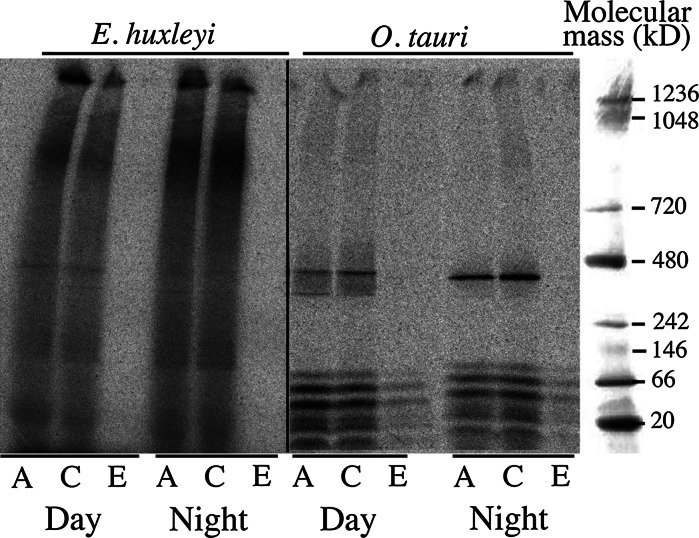
Fig. 6.
Autoradiography of dried gels after separation of whole cell extracts on blue native PAGE. O. tauri and E. huxleyi cells were grown for 5 d under standard conditions (Mf medium+0.1 μM ferric citrate) and a 12:12 light–dark regime. Cells in exponential growth phase were harvested in the middle of the day (“Day”) or in the middle of the night (“Night”), washed once by centrifugation with iron-free Mf medium and incubated in the same medium for 2.5 h (E. huxleyi) or 1.5 h (O. tauri) in the light at 20 °C with either 2 μM 55ferrous ascorbate (1:100; “A”), 2 μM 55ferric citrate (1:20; “C”) or 2 μM 55ferric EDTA (1:20; “E”). Cells were then washed once by centrifugation with iron-free Mf medium (E. huxleyi) or (O. tauri) with a medium containing strong iron chelators (see Sect. 2), and whole cell extracts were prepared by sonication. After native PAGE (about 25 μg protein per lane), the gels were dried and autoradiographed

