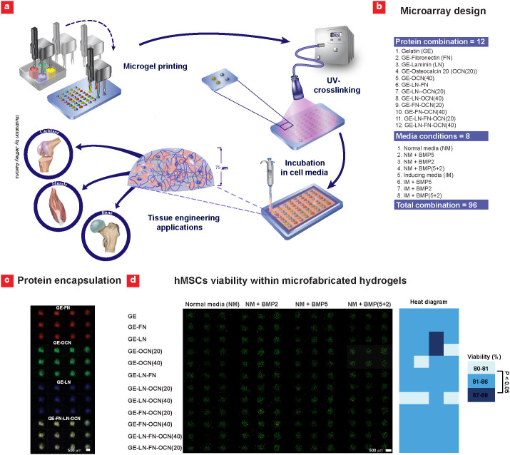
Figure 1. Fabrication of 3D hMSC-laden gel microarray.
(a) A robotic microarray spotter was used to rapidly print droplets consisting of hMSCs, gelatin methacrylate (GE)-based prepolymer solution and various ECM proteins on TMSPMA functionalized glass slide. The printing step was followed by a 15 sec UV light exposure to form the miniaturized cell-laden constructs. Following printing, cell-laden gel microarrays were placed inside sealed chambers (Illustration made by Jeffrey Aarons). (b) Various combinations of ECM proteins and media formulations were used to conduct the microarrays experiments. The concentration of LN and FN was selected to be 40 μg/ml while OCN was printed at two concentrations of 20 μg/ml and 40 μg/ml. (c) Fluorescence images of the encapsulated proteins within the hydrogel constructs after 24 hours in solution. (d) hMSCs viability within 48 combinatorial 3D microenvironments in normal (control) media after 7 days of culture along with color-diagram displaying the quantified cell viability (n = 3–9).

