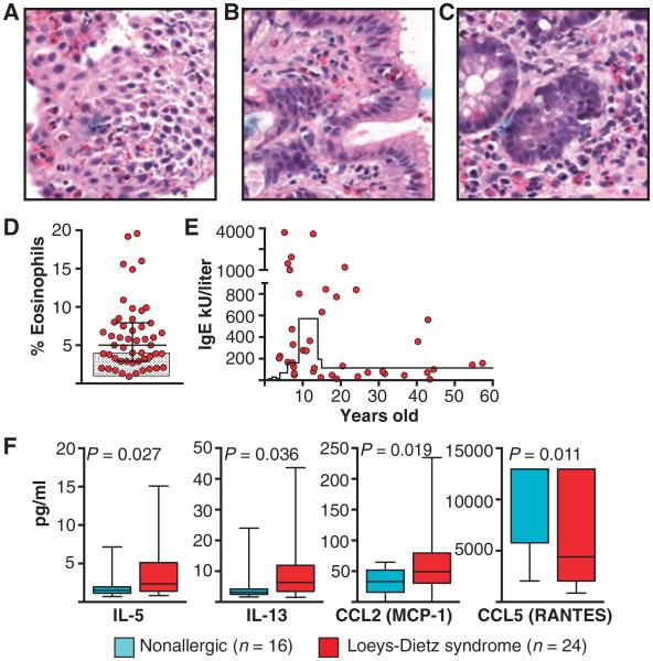
Fig. 1. Evidence for allergic predilection in LDS patients.
(A to C) Biopsies stained with hematoxylin and eosin demonstrating an eosinophilic infiltrate in the esophagus (A), stomach (B), and colon (C) of a child with LDS. Magnification, ×40. (D) Percentage of eosinophils in the peripheral blood of LDS patients (n = 50). Levels were significantly increased (P = 0.009) compared to the norm (shaded box) by Wilcoxon test. Line and whiskers indicate mean and SD, respectively. (E) Total serum levels of IgE (kU/liter) from LDS patients versus age (n = 41). Levels were elevated (P = 0.016; Student’s t test, two-tailed) above the 95% confidence interval for age as indicated by the solid line. Each point represents an individual patient in (D) and (E). (F) Levels of IL-5, IL-13, CCL2 (MCP-1), and CCL5 (RANTES; pg/ml) in plasma from patients with LDS (n = 24) and age-matched nonallergic controls (n = 16). Significant P values are indicated; comparisons were done by Wilcoxon test.

