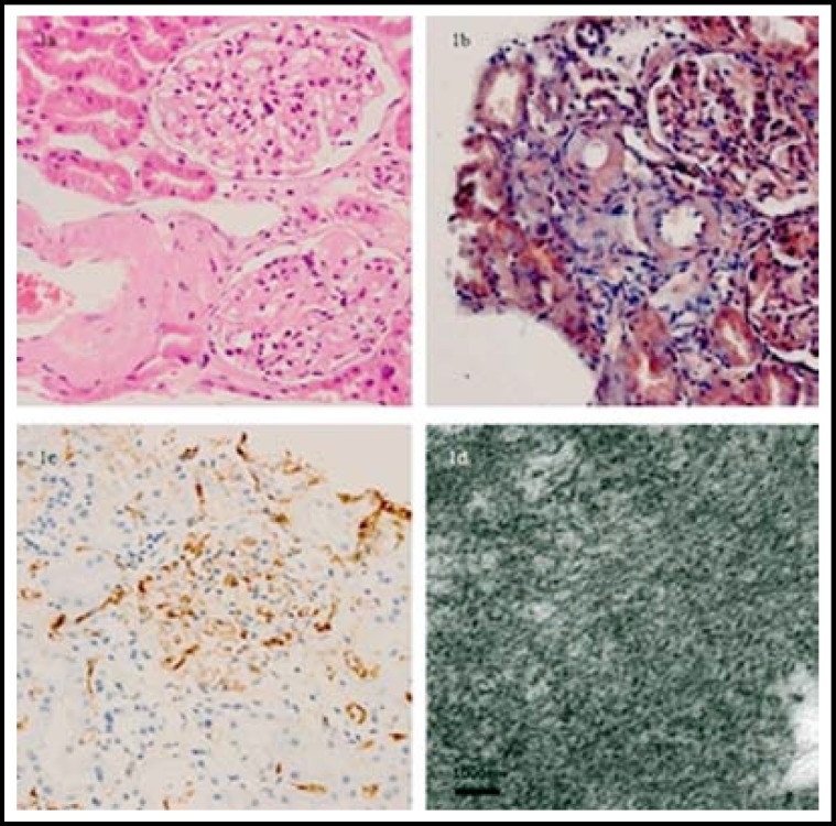
Fig.1.
Histopathological findings of the renal biopsy in a 46-year-old man with systemic amyloidosis. Amyloid deposition in the interstitial of kidney demonstrated (a) red by HE staining (×200) and (b) orange by Congo red staining (× 200). The interstitial material is positively stained by Lambda staining and showed (c) brown (× 200). (d) Deposits composed of nonbranching fibrils were showed under the electron microscopy (× 8000).

