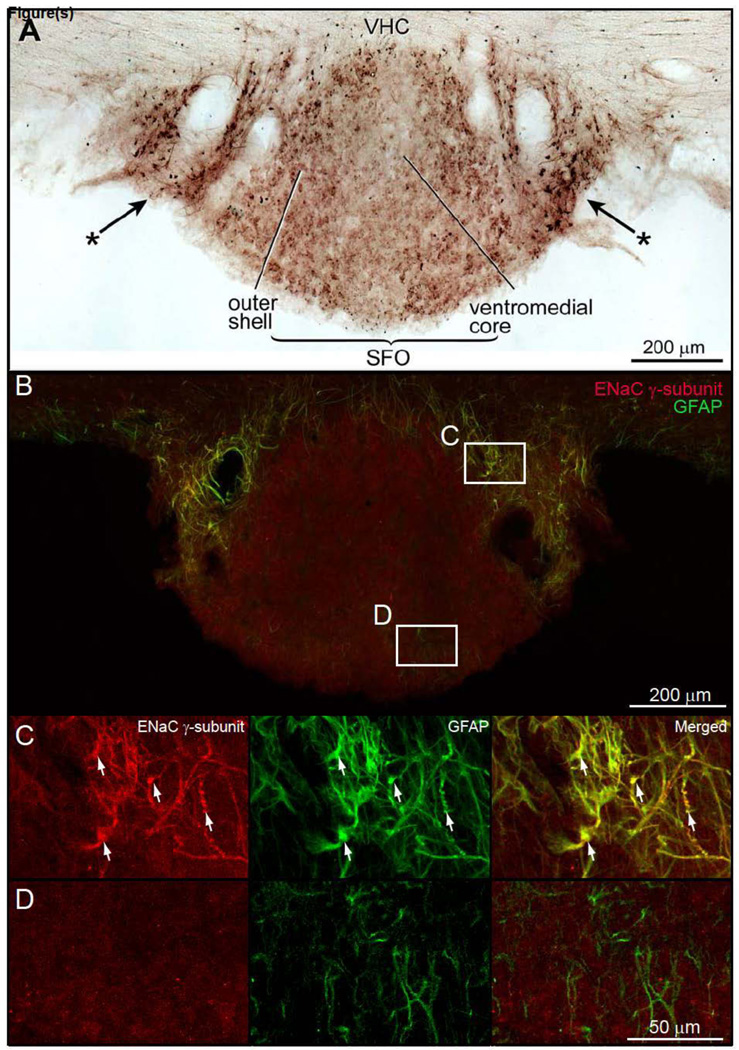
Figure 3.
Brightfield preparation of a transverse section through the subfornical organ (SFO) to demonstrate ENaC γ-immunoreactive neurons and astrocytes. A. Very weak ENaC γ-expressing neurons were seen in both the ventromedial core and outer shell of the SFO. Intensely immunostained ENaC γ-positive astroglia were present on the lateral borders of the SFO (*→). B. Double immunofluorescence preparation to show ENaC γ-expressing astrocytes in the lateral border of the SFO. The expression of ENaC γ-subunit in the ventromedial core and outer shell of the SFO is much lower than the astrocytes in the border zone. In this particular preparation, ENaC γ-subunit immunoreactivity in neurons was below detectable levels. Other preparations (data not shown) show ENaC γ-subunit and NeuN co-localization, indicating that SFO neurons express low levels of ENaC γ-subunit. C. In the SFO border zone, astrocytes co-express ENaC γ-subunit protein and GFAP. D. GFAP immunoreactive astrocytes in the core of the SFO do not co-contain ENaC γ-subunit immunoreactivity.

