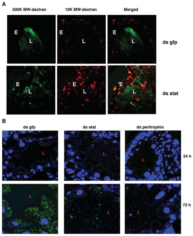
Figure 6. Borrelia burgdorferi leaves the gut lumen and attaches to the gut epithelial cells during colonization.
Confocal microscopy of: A. 24 h fed guts of ds gfp or ds stat RNA-injected nymphs that were capillary-fed Rhodamine red-conjugated 10,000 molecular weight dextran (10 K MW dextran) and Fluorescein-conjugated 500,000 molecular weight dextran (500 K dextran). Magnification x40. ‘L’ marks the lumen and ‘E’ the gut epithelium. B. Carnoy’s fixed and sectioned guts from dsgfp, dsstat, or dsperitrophin RNA-injected nymphs fed for 24 and 72 h on B. burgdorferi-infected C3H mice. Midgut nuclei and spirochetes stained with TO-PRO-3 (blue) and FITC-conjugated B. burgdorferi antisera (green), respectively. Magnification x63. ‘L’ marks the gut lumen.

