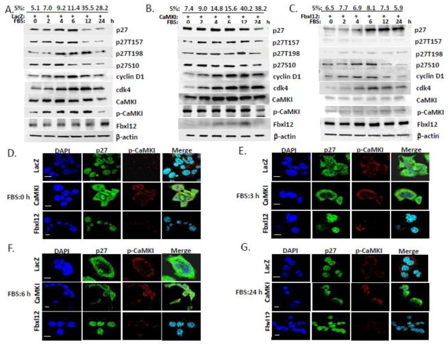
Figure 4. CaMKI controls cell cycle progression in lung epithelia via p27 phosphorylation and its subcellular localization.
A549 cells were transfected with LacZ (A), CaMKI (B) or Fbxl12(C) plasmids followed by arrest at G0 by starvation. In 48 h 10% FBS was added and cells were collected at indicated times for flow cytometry and immunoblot analysis. The percentage of cells that entered into the S phase of the cell cycle are indicated at the top (S%). (D–G) CaMKI regulates p27 localization. MLE cells transfected with LacZ, CaMKI or Fbxl12 as described above were fixed and processed for immunostaining using p-CaMKI and p27 antibodies at indicated times. DAPI was used to visualize the nucleus. Scale bar, 10 μm. The data from each panel represents at least n=3 separate experiments.

