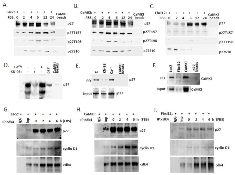
Figure 5. CaMKI binds p27 and regulates cdk4/cyclinD1/p27 complex formation.
CaMKI binds p27. (A–C) A549 cells transfected with LacZ (A), CaMKI (B) and Fbxl12 (C) were starved for 48 hours and then recovered with addition of 10% FBS. Cells were then collected and processed for pull down assays using CaMKI-conjugated beads followed by immunoblotting with appropriate antibodies. (D) Direct binding between CaMKI and p27. Purified recombinant p27 was pre-incubated with CaMKI beads with or without Ca2+ or KN-93 as indicated on top of the lanes, followed by immunoblot analysis with p27 antibody. (E) Cells exposed to KN-93 (20μM) or Ca2+(2mM) for 1 h were then collected followed by pull down assays using CaMKI-conjugated beads. (F) Cells transfected as described above (A–C) were processed for pull down assays using beads conjugated to p27 followed by immunoblot analysis with CaMKI antibody. (G–I) CaMKI regulates cdk4/cyclinD1/p27 complex formation. Cells transfected with LacZ (G), CaMKI (H) or Fbxl12 plasmids (I) were released with 10% FBS addition and then collected at indicated times for immunoprecipitation using cdk4 antibody. Immunoprecipitants and input were analyzed by imuunoblotting using p27 and cyclin D1 antibodies. n=2 experiments.

