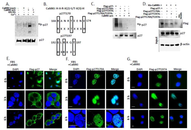
Figure 7. CaMKI phosphorylates mouse p27 and regulates its intracellular localization in mouse lung epithelium.
(A) Purified CaMKI phosphorylates p27 purified from mouse cells in vitro. Purified p27 from MLE cells was incubated with 10 μCi [γ-32P]ATP in the presence or absence of an active or heat-inactivated form of CaMKI (500 nM) and Ca2+. After 1 h of incubation with or without KN-93 at 30°, reactions were terminated with 4X Laemmli protein loading buffer and products were resolved by 10% SDS-PAGE and analyzed by autoradiography and immunoblotting using p27 antibodies. (B) Alignment of the CaMKI consensus recognition sequence and putative phosphorylation sites within mouse p27. (C) CaMKI phosphorylates p27 WT but not mutants in mouse lung epithelia. MLE cells were transfected with Flag-tagged p27 WT or mutants p27T170A, p27T197A or p27T170A / T197A. Flag-tagged proteins were then pulled down with Flag-agarose. The Flag-p27-, Flag-p27T157A-, Flag-p27T198A- and Flag p27T170A / T197A -beads were dephosphorylated using alkaline phosphatase and then incubated with 10 μCi [γ-32P]ATP in the presence of an active form of CaMKI (500 nM) and Ca2+ (2 mM). After 1 h of incubation at 30°, reaction products were resolved by 10% SDS-PAGE and analyzed by autoradiography and immunoblotting. (D) CaMKI associates with p27 wild type but not with point mutants. MLE cells were co-transfected with His-CaMKI and either Flag-p27, Flag-p27T157A, Flag-p27T198A or Flag-p27T170A / T197A plasmids followed by pull down with His-conjugated beads. Samples were then analyzed by immunoblotting using protein specific antibodies. (E–G) MLE cells were co-transfected with CaMKI and either Flag-p27 (E), Flag-p27T170A (F) or Flag-p27T197A plasmids (G). Cells were then synchronized by serum starvation and released in 48 h by addition of 10% FBS followed by fixation at indicated times and immunostaining using anti-Flag antibody and DAPI to visualize nuclei. The data are from n=3 experiments. Scale bar, 10 μm.

