Figure 7.
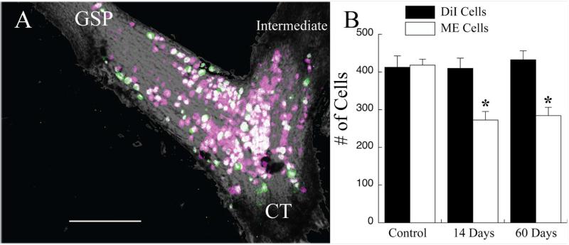
A: Photomicrograph of an image stack showing geniculate ganglion cells that were labeled with DiI at the time of the CTX (magenta cells) and those that were labeled with micro-emerald when the nerve was labeled 60 days later (green cells). Magenta-only cells are cells in which peripheral axons were not labeled following the CTX. Double-labeled cells (white cells) are neurons that had peripheral axons at both nerve labeling times. There were no green-only labeled cells. This is a collapsed image; therefore, some cells may obscure the visualization of others. Data were collected from single optical sections. GSP, greater superficial petrosal nerve; CT, chorda tympani nerve; intermediate, intermediate nerve. B: Mean (±SEM) number of cells labeled with DiI (solid bars) and with micro-emerald (open bars) in control rats or in rats with a CTX 14 or 60 days before labeling of the chorda tympani nerve with micro-emerald. There was no change in the number of DiI-labeled geniculate ganglion cells post-CTX; however, the number of micro-emerald cells significantly decreased by 14 days post-CTX. *P < 0.05 compared with the control group. Scale bar = 200 μm.

